|
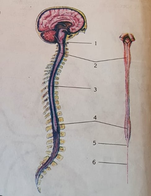
- in the vertebral canal = the spinal canal (with adipose tissue), formed by the overlapping of the vertebral orifices, which it does not occupy entirely. - the upper limit of the spinal cord corresponds to the occipital hole or the emergence of the first spinal nerve (it goes above I cervical vertebrae, C1), and the lower limit is in front of the L2 vertebra - under the L2 vertebra, the spinal cord extends with the medullary cone, and it with the terminal phylum - on either side of the medullary cone and phylum is the ponytail with an almost vertical direction, formed by the lumbar and sacral nerves Terminal phylum + sacral and lumbar nerves (L3, L4 - L5), form the ponytail. - cylindrical shape, slightly flattened antero-posteriorly
- located centrally, in the form of letters H or columns with a longitudinal arrangement and horns with a transverse arrangement. -consisting of the bodies of neurons, so here are the nerve centers. -The H-bar forms the gray commissure of the spinal cord which presents the ependymal canal with cerebrospinal fluid. -the part of the gray commissure anterior to the ependymal canal is called the anterior gray commissure and the part of the gray commissure posterior to the ependymal canal is called the posterior gray commissure
- between the anterior gray commissure and the anterior median fissure is the anterior white commissure -between the posterior gray commissure and the posterior median groove is the posterior white commissure - consisting of extensions of neurons (axons grouped in bundles = ropes = cords) - has 3 pairs of cords - rear, side, front - Cords are conduction paths = pulse tracts between different stages of the CNS. 3 pairs of horns:
made by gray matter by somatic and vegetative neurons
performed by white matter through cords = bundles = tracts